Imaging and STED
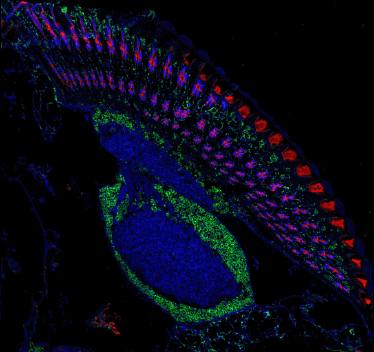
The essential feature of confocal microscope is a diapharagm, called a pinhole, that blocks the light emitted from points lying outside the focal plane. This results in exceptional sharpness, contrast and resolution of the confocal images. In addition, the use of a beam of coherent light enables selective excitation of specific fluorophores, while point scanning allows three-dimensional reconstruction of the specimen volume (Fig. 1). Accordingly, the confocal microscopy forms another dimension of microsocpy. The Nencki Institute is equipped with two high-end confocal systems (see http://konfokal.nencki.gov.pl/).
In spite of its superior quality, confocal imaging is bound by the same basic resolution limit as the conventional light microscopy, being unable to resolve objects that are smaller than about 250 nm. This is known as the Abbe’s limit, derived from the diffraction properties of the light. It is in fact a big problem for cell biological studies because the dimensions of many important organelles, ranging between 10 and 200 nm, make them invisible or visible as
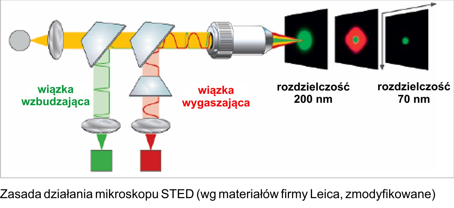
fuzzy blobs only. Nencki Institute has recently got an advanced confocal system (Leica TCS SP5) equipped with a superresolution STED (stimulated emission depletion) attachment that allows optical imaging with the resolution beyond the diffraction limit. STED was invented by German physicist Stefan Hell (http://www.mpibpc.mpg.de/groups/hell/). Compared to conventional confocal, STED utilizes, in addition to an excitation beam, a second doughnut-
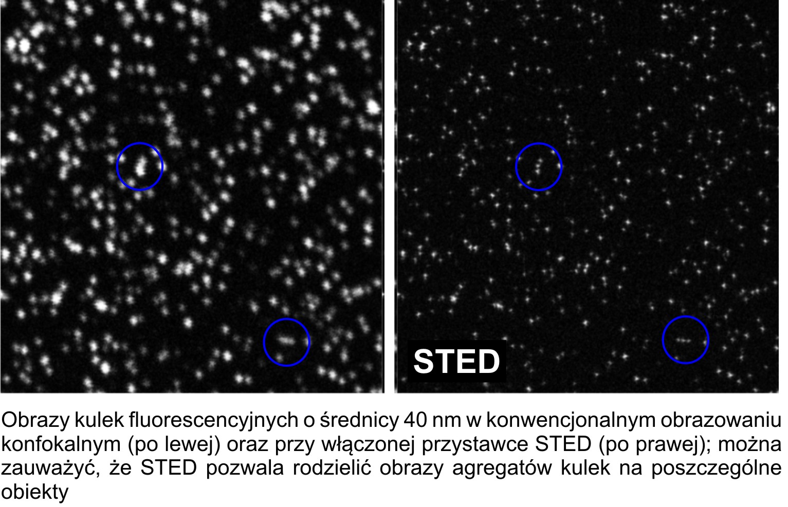
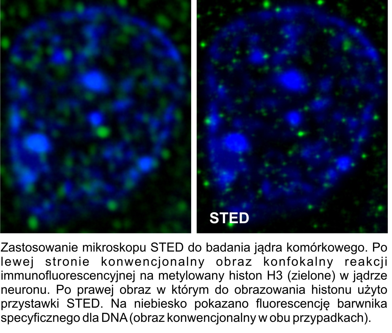
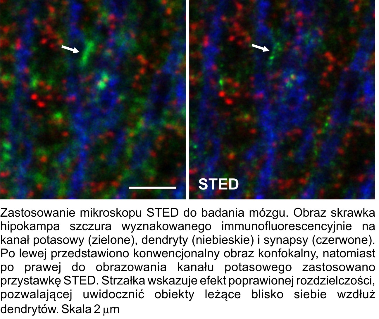
shaped laser beam that depletes the fluorescence in the outer rim of an excited spot (Fig. 2A). This results in a considerable improvement in lateral resolution. The STED system from Nencki Institute allows imaging with a resolution of 70 nm, as measured using fluorescent beads (Fig. 2 C, D). Although at present commercial STED systems have only a single STED channel, the other channels can be imaged sequentially with a conventional resolution, enabling the comparison of superresolved structures to other elements of cells and tissues (Fig. 2 E-K).
Date
06 July 2010
Gallery