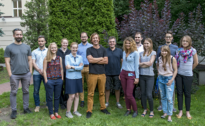
- Head of laboratory
- Scientific Staff
- Technician and administration staff
- PhD Students
- Research profile
- Current research activities
- Selected publications
Head of laboratory

Research profile
Laboratory of Brain Imaging (LOBI) is one of the core facilities at the Neurobiology Center. LOBI provides access to state-of-the-art research support and technologies for internal and external scientists. Technologies used at LOBI include: magnetic resonance imaging (MRI), spectroscopy (MRS), electroencephalography (EEG – including EEG-fMRI simultaneous recordings), transcranial magnetic stimulation (TMS) and computational image analysis.
Research at LOBI has made fundamental contributions to the understanding of the neural processes responsible for cross-modal neuroplasticity in healthy and deaf people, neuronal mechanisms of consciousness, processing of emotionally charged information in healthy and subclinical populations as well as analytic approaches for multisite MRI data of developmental and neurological disorders.
Additionally, LOBI develops standardized databases of stimuli to facilitate highly controlled experimental research on emotion and cognition – Nencki Affective Picture System (1356 images) and Nencki Affective Word datasets (2902 Polish words). Stimuli in both datasets are characterized in terms of emotional valence, arousal, as well as basic emotion intensities. The NAPS and NAWL databases are freely accessible to the scientific community (http://exp.lobi.nencki.gov.pl/dnaps). More information can be found on the web page: http://lobi.nencki.gov.pl/
Current research activities
• Brain plasticity in language acquisition. The aim of the project is to reveal a multimodal brain network for speech and print in a second language. Adopting a longitudinal design, we test learners of foreign languages, sign language and Braille reading. Besides distin- guishing which structures of the language-processing network are multimodal and which are not, we plan to examine the dynamic changes in the brain morphology and function associated with linguistic learning.
• Neuronal mechanisms of consciousness. We develop two lines, first we study how brain activity differs between unconscious (e.g. sleep, anesthesia) and conscious states. Second, we try to address which mechanisms allow external stimuli to gain access to our subjective “stream of consciousness”.
• Neural correlates of procrastination. Using cognitive tasks in emotional contexts during fMRI experiments we try to get neuronal-level insight into mechanisms of this self-regulatory failure.
• Influence of emotions of long-term and associative memory
• Directed forgetting of emotionally charge material using NAWL
• Neural markers of dyslexia.
Selected publications
Gola M., Wordecha M., Sescousse G., Lew-Starowicz M., Kossowski B., Wypych M., Makeig S., Potenza M.N., Marchewka A. (2017) Can pornography be addictive? An fMRI study of men seeking treatment for problematic pornography use. Neuropsychopharmacology, doi:10.1038/npp.2017.78.
Bola Ł., Zimmermann M., Mostowski P., Jednoróg K., Marchewka A., Rutkowski P., Szwed M. (2017) Task-specific reorganization of the auditory cortex in deaf humans. Proc Natl Acad Sci USA, 114(4): E600-E609.
Płoński P., Gradkowski W., Altarelli I., Monzalvo K., van Ermingen-Marbach M., Grande M., Heim S., Marchewka A., Bogorodzki P., Ramus F., Jednoróg K. (2017) Multi-parameter machine learning approach to the neuroanatomical basis of developmental dyslexia. Human Brain Mapp, 38(2): 900-908.
Michałowski J.M., Matuszewski J., Droździel D., Koziejowski W., Rynkiewicz A., Jednoróg K., Marchewka A. (2016) Neural response patterns in spider, blood-injection-injury and social fearful individuals: new insights from a simultaneous EEG/ECG–fMRI study. Brain Imaging Behav, doi:10.1007/s11682-016-9557.
Marchewka A., Kherif F., Krueger G., Grabowska A., Frąckowiak R., Dragański B. (2014) Influence of magnetic field strength and image registration strategy on voxel-based morphometry in a study of Alzheimer’s disease. Human Brain Mapp, 35(5): 1865-1874.

